Why Your Knee X-ray Doesn’t Tell the Whole Story: The Science of Joint Preservation

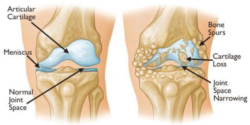
If you have knee pain, an X-ray is often one of the first investigations. It can show joint-space narrowing, bone spurs and other signs of arthritis. But it does not show cartilage directly: the space between the bones is used as an indirect indication of cartilage loss.

An X-ray is valuable, but it cannot tell us everything about an individual knee. It cannot show how much pain you feel, directly show the joint lining or menisci, or reveal bone marrow lesions in the way an MRI can. People with similar X-ray findings can have different symptoms and levels of function. [1]

That does not mean an X-ray is unimportant—or that knee replacement is the wrong choice. It means the image should be considered alongside your symptoms, examination, activity goals and other relevant findings.

Pain can involve several tissues

Articular cartilage itself has no nerve endings. Pain in an arthritic knee can involve other tissues, including the synovium, subchondral bone, menisci and supporting structures. MRI studies have found associations between knee pain and findings such as bone marrow lesions, synovitis or effusion, and meniscal changes. These associations do not prove which finding is causing pain in any one person. [2]

An MRI can help identify features that an X-ray does not show. It is not needed for every person, and an MRI finding must still be interpreted in the context of the history and examination.

The role of inflammation and metabolic health

Osteoarthritis affects more than cartilage and includes biological and inflammatory processes. The degree and sources of inflammation differ between people.

Blood tests may be considered as part of an individual assessment. For example, HbA1c can help assess longer-term blood sugar control. High-sensitivity C-reactive protein (hs-CRP) is a nonspecific marker in the blood; it does not locate inflammation in the knee or identify a single cause of arthritis. Research has not established hs-CRP as the root cause of knee osteoarthritis. [3]

Where a health issue needs attention before a treatment is considered, the plan may include addressing it first. The evidence does not establish that a particular blood-test result predicts how an individual will respond to a biological treatment.

How we assess whether knee preservation is appropriate

At the Madras Joint Rejuvenation Centre, assessment considers three connected areas:

Area What we consider
The Joint Symptoms, examination, joint surfaces, menisci, synovium and stability
The Bone The bone beneath the cartilage, including MRI findings such as bone marrow lesions when imaging is indicated
The Person Overall health, strength, activity demands, goals and ability to complete rehabilitation

At MJRC, PRASAD (Platelet Rich Adipose Stromal Advanced Derivative) describes an individualized joint-preservation framework. The word “Advanced” is an open slot: the treatment plan is selected according to the patient’s findings. It is not a fixed recipe, and not every person receives every biological component.

Depending on the assessment, selected orthobiologic approaches may include low-leukocyte platelet-rich plasma, adipose-derived stromal vascular fraction prepared by fractionation, or bone marrow aspirate concentrate. The proposed target and method of delivery depend on the pathology being addressed.

Biological treatment cannot guarantee cartilage regrowth, a permanent cure, or avoidance of knee replacement. Some selected patients may experience less pain and better function, but results vary and the evidence differs across treatments.

For a closer look at the research on bone marrow lesions and the bone beneath the cartilage, read The Bone Beneath the Cartilage: What the Research Shows.

Your next step

A knee-preservation decision requires more than an X-ray grade. It brings together your symptoms, a physical examination, appropriate imaging and relevant aspects of your overall health and goals.

A full assessment may find that preservation is reasonable, or that knee replacement is the more appropriate option. Discuss the benefits, limitations and alternatives with an orthopaedic surgeon before making a decision.

About the author

Dr. A.K. Venkatachalam, MS (Orth), DNB (Orth), FRCS (Glasg), MCh (Orth) (Liverpool), is the founder and chief orthopaedic surgeon at the Madras Joint Rejuvenation Centre in Chennai. He pioneered microfracture surgery in Chennai in 2004 and autologous chondrocyte implantation in 2014. His clinical practice focuses on biological joint preservation and orthobiologics.

TNMC Reg. No.: 38088

References

  1. Neogi T, et al. Association between radiographic features of knee osteoarthritis and pain: results from two cohort studies. BMJ. 2009;339:b2844. doi:10.1136/bmj.b2844.
  2. Wang X, et al. Synovitis mediates the association between bone marrow lesions and knee pain in osteoarthritis: data from the Foundation for the National Institute of Health (FNIH) Osteoarthritis Biomarkers Consortium. Osteoarthritis Cartilage. 2022;30(9):1270–1277. doi:10.1016/j.joca.2022.06.004.
  3. Funck-Brentano T, et al. Causal factors for knee, hip, and hand osteoarthritis: a Mendelian randomization study in the UK Biobank. Arthritis Rheumatol. 2019;71(10):1634–1641. doi:10.1002/art.40928.

Disclaimer: This article is for education and does not replace an individual medical assessment. Treatment benefits and risks vary. No treatment described here guarantees healing, cartilage restoration or avoidance of surgery.

The Bone Beneath the Cartilage: What the Research Shows

As of 7 November 2026 · Dr. A.K. Venkatachalam

Why the bone matters

Published research now supports treating the bone beneath the cartilage as a target in its own right. Bone marrow lesions change over time, raised pressure inside the bone is linked to deep pain at rest, and two controlled trials show that treatment delivered into the bone does better than the same treatment without it.

Articular cartilage has no nerves. The subchondral bone beneath it does, along with blood vessels and a marrow space that remodels throughout life. On MRI, the abnormal areas in this bone are called bone marrow lesions (BMLs); reports often call them bone marrow oedema.

This page summarises the published evidence behind the subchondral component of the PRASAD framework. Every study cited here was carried out by other investigators. Each section is tagged with the kind of evidence it rests on.

Bone marrow lesions are not fixed

Evidence: human, longitudinal cohort, MRI (natural history, no treatment)

In the Multicenter Osteoarthritis Study, 395 knees were scanned twice, about 30 months apart. Two-thirds (66%) of the lesions present at the start had changed in size, and half had shrunk or disappeared (Roemer et al., 2009).

The direction of change mattered. Lesions that grew, and new lesions, carried a much higher risk of cartilage loss in the same region of the knee.

A bone marrow lesion is therefore an active process, not a permanent scar. That is what makes it a reasonable target for treatment.

Why the bone hurts: pressure inside a rigid compartment

Evidence: human, direct pressure measurement and perfusion MRI (mechanism)

In 1975, Arnoldi and colleagues measured pressure inside the femur and tibia near the knee in 53 patients. Pressure was low in patients with neither arthritis nor rest pain, and in half of those with arthritis but no rest pain; aching rest pain went with raised intraosseous pressure (Arnoldi et al., 1975). In a later series, every patient with typical rest pain showed venous stasis and raised pressure in the marrow near the painful joint (Arnoldi et al., 1980).

The mechanism follows from the anatomy. Blood keeps entering the bone, but its drainage is obstructed. Bone cannot expand, so the pressure must rise until blood is forced out through the narrowed exits.

Modern imaging agrees. Dynamic contrast MRI in 15 osteoarthritic knees and 7 controls showed reduced venous outflow and delayed clearance in the subchondral bone, consistent with venous stasis (Aaron et al., 2018).

What the bright MRI signal shows. On fat-suppressed sequences such as STIR, normal fatty marrow goes dark, so the bright area is water. In clinical interpretation that fluid has two sources: passive (venous congestion that cannot drain) and active (leakage from inflamed capillaries). Tissue examined after knee replacement shows necrosis, fibrosis and remodelled trabeculae within these zones; free fluid is largely lost when tissue is processed (Zanetti et al., 2000). The fluid is the active process; the scarring is what it leaves behind.

What goes into the bone matters

Evidence: human, randomised, double-blind, multicentre controlled trial

Eighty-six patients with Kellgren–Lawrence grade III–IV knee osteoarthritis were randomised. Both groups received the same intraosseous procedure followed by three intra-articular injections of plasma rich in growth factors (PRGF). The only difference was what went into the bone: PRGF in one group, saline in the other (Sánchez Santiuste et al., 2025).

The intraosseous PRGF group improved significantly more in pain, function and quality of life across most time points to one year. No serious adverse events were recorded.

Because both groups had the needle in the bone and the same joint treatment, the trial isolates the biological material delivered into the bone. It did not include follow-up MRI, so it shows a clinical effect, not lesion regression.

Disclosure: one co-author, E. Anitua, developed the PRGF system.

Where it goes matters

Evidence: human, randomised within-patient trial, 13–18 years’ follow-up

Sixty patients with comparable osteoarthritis in both knees had bone marrow concentrate harvested once and divided equally. After randomisation, one knee received it into the subchondral bone of the femur and tibia; the other received it into the joint (Hernigou et al., 2021).

At two years, clinical and MRI improvement was greater on the subchondral side. At an average of 15 years:

Delivery route Knees Went on to knee replacement
Subchondral bone 60 12 (20%)
Joint (intra-articular) 60 42 (70%)

The same patient, the same cells and the same dose were on both sides; only the route differed. That removes metabolism, lifestyle and general health as explanations for the gap.

Treating the lesion directly

Evidence: human, retrospective series with MRI; systematic review and meta-analysis

Subchondral bone marrow concentrate. In a retrospective analysis of 62 patients with knee osteoarthritis and MRI-confirmed bone marrow lesions, bone marrow aspirate concentrate (5 mL per site) was injected into the affected subchondral bone. Repeat MRI at 3 months showed reduced lesion scores, and pain and function improved through 12 months of clinical follow-up (Ezzi et al., Journal of Orthopaedic Surgery and Research, 2026). The study had no control group, so it documents improvement after treatment rather than isolating the injection’s effect.

Subchondroplasty. A different approach fills the lesion with calcium phosphate bone substitute for mechanical support. A 2026 meta-analysis of 12 studies and 829 patients found clinically important improvement in pain and function from one month to at least two years, with 2.4% converted to knee replacement at one year (Klincke et al., 2026). The authors note short follow-up and a lack of control groups.

The two approaches work differently, one biological and one mechanical, and neither establishes the other’s mechanism. Both show the bone beneath the cartilage can be treated.

What this establishes, and what it does not

Taken together, studies by different groups, with different methods and patients, converge on one conclusion: the subchondral bone takes part in the disease and can be treated.

Established

  • Bone marrow lesions change over time, and their course tracks cartilage loss (MOST).
  • Raised intraosseous pressure and venous stasis accompany deep pain at rest (Arnoldi; Aaron).
  • Changing what is delivered into the bone changes the clinical result, under double-blind conditions (Sánchez Santiuste).
  • Changing where the same cells are delivered changes long-term replacement rates in the same patient (Hernigou).

Not yet established

  • That shrinking a lesion means cartilage has regrown.
  • That every lesion needs treatment, or that all lesions share one cause.
  • A consistent benefit in every study: a case-matched registry comparison found no added benefit in patient-reported outcomes from adding intraosseous bone marrow concentrate and platelets to joint injection (Centeno et al., 2021). It was not randomised, and its authors noted a possible benefit in large lesions that the sample was too small to confirm.

How this informs PRASAD

PRASAD (Platelet Rich Adipose Stromal Advanced Derivative) is a treatment framework that combines selected orthobiological approaches according to the pathology in each knee. It treats the joint and the bone beneath it as distinct targets, which is what the evidence above supports.

  • The joint: leukocyte-poor platelet-rich plasma and adipose-derived stromal tissue, delivered into the joint.
  • The bone: bone marrow aspirate concentrate delivered into the subchondral bone, as a standing part of the framework.
  • The open slot: the “Advanced” in the name is deliberately open, so the framework can take in what further evidence supports. A-Kavach™ (an autologous alpha-2-macroglobulin fraction) is its first occupant.

Treatment follows assessment: imaging, metabolic health, alignment, muscle function and the patient’s readiness for rehabilitation.

The studies on this page did not test the full PRASAD™ framework. They support its subchondral component. Outcomes in our own patients are being documented systematically, and will be reported as they mature.

References

  1. Roemer FW, Guermazi A, Javaid MK, et al. Change in MRI-detected subchondral bone marrow lesions is associated with cartilage loss: the MOST Study. Ann Rheum Dis. 2009;68(9):1461–1465. doi:10.1136/ard.2008.096834
  2. Arnoldi CC, Lemperg RK, Linderholm H. Intraosseous hypertension and pain in the knee. J Bone Joint Surg Br. 1975;57-B(3):360–363. doi:10.1302/0301-620X.57B3.360
  3. Arnoldi CC, Djurhuus JC, Heerfordt J, Karle A. Intraosseous phlebography, intraosseous pressure measurements and 99mTc-polyphosphate scintigraphy in patients with various painful conditions in the hip and knee. Acta Orthop Scand. 1980;51:19–28. doi:10.3109/17453678008990764
  4. Aaron RK, Racine JR, Voisinet A, Evangelista P, Dyke JP. Subchondral bone circulation in osteoarthritis of the human knee. Osteoarthritis Cartilage. 2018;26(7):940–944. doi:10.1016/j.joca.2018.04.003
  5. Zanetti M, Bruder E, Romero J, Hodler J. Bone marrow edema pattern in osteoarthritic knees: correlation between MR imaging and histologic findings. Radiology. 2000;215(3):835–840. doi:10.1148/radiology.215.3.r00jn05835
  6. Sánchez Santiuste M, Vaquerizo García V, Pareja Esteban JA, Prado R, Padilla S, Anitua E. PRGF versus saline intraosseous infiltrations combined with intra-articular PRGF in severe knee osteoarthritis: a prospective double-blind multicentric randomized controlled trial with 1-year follow-up. J Clin Med. 2025;14(22):8075. doi:10.3390/jcm14228075
  7. Hernigou P, Bouthors C, Bastard C, et al. Subchondral bone or intra-articular injection of bone marrow concentrate mesenchymal stem cells in bilateral knee osteoarthritis: what better postpone knee arthroplasty at fifteen years? A randomized study. Int Orthop. 2021;45(2):391–399. doi:10.1007/s00264-020-04687-7
  8. Ezzi SH, Ding H, Wu S, et al. Subchondral bone marrow aspirate concentrate injection improves clinical outcomes and reduces bone marrow lesions in knee osteoarthritis: 12-month retrospective analysis. J Orthop Surg Res. 2026.
  9. Klincke V, Demoor B, Dehouwer F, Tampere T, et al. Subchondroplasty for bone-marrow lesions in the osteoarthritic knee improves pain and function: a systematic review and meta-analysis. The Knee. 2026. doi:10.1016/j.knee.2026.104441
  10. Centeno C, Cartier C, Stemper I, et al. The treatment of bone marrow lesions associated with advanced knee osteoarthritis: comparing intraosseous and intraarticular injections with bone marrow concentrate and platelet products. Pain Physician. 2021;24(3):E279–E288. PubMed 33988949

Dr. A.K. Venkatachalam · MS (Orth), DNB (Orth), FRCS (Glasg), MCh (Orth) (Liverpool) · Tamil Nadu Medical Council Reg. No. 38088 · Madras Joint Rejuvenation Centre, Kasthuri Hospital, West Tambaram, Chennai

General health education. Individual clinical assessment is necessary before treatment decisions.

A-Kavach– The Biological Shield That Protects Your Joints From Arthritis

A-Kavach™ – The Biological  shield That Protects Your Joints From Arthritis

Arthritis is not just “wear and tear.” For decades, patients were told their cartilage simply eroded because of aging or excessive load. But modern joint biology tells a more complete story:

**Your knee is under continuous chemical attack.**

Inside an arthritic joint, destructive enzymes, inflammatory molecules, and oxidative stress work like microscopic knives, cutting through cartilage and accelerating its breakdown — often long before your X-ray shows severe change. That’s why some patients with only mild X-ray findings have significant pain, and some with advanced-looking X-rays have very little.

**A-Kavach™** is designed to interrupt that chemical attack — as the first step in a two-part biological strategy.

—

## Why “Wear and Tear” Alone Doesn’t Explain Arthritis

For years, osteoarthritis was treated as a purely mechanical problem — a hinge wearing down. But we now understand:

– Cartilage breakdown is driven substantially by biochemical inflammation
– Enzymes such as MMPs and ADAMTS, along with inflammatory cytokines, break down collagen and proteoglycans in cartilage
– This process can begin long before it’s visible on an X-ray
– Mechanical load is a contributing factor — but the chemical cascade is what drives ongoing destruction

A-Kavach™ is built specifically to interrupt that chemical cascade — before more cartilage is lost.

What Is A-Kavach™?

A-Kavach™ is an autologous (from your own blood) biological shield developed at the **Madras Joint Rejuvenation Centre (MJRC)**, Kasthuri Hospital, Chennai. It is concentrated from your own platelet-poor plasma and is rich in alpha-2 macroglobulin (A2M) — a naturally occurring protein that neutralizes the destructive enzymes driving cartilage breakdown.

**A-Kavach™ is the first step, injected before anything else, because its protective action needs to take effect immediately.** Once the joint’s chemical environment is no longer actively hostile, we follow with the **PRASAD™ Protocol** — our platelet-rich, stromal, and marrow-derived biologic combination — to work within a joint that is no longer under active biochemical attack.

*(You can read the full explanation of the PRASAD™ Protocol here →
A-Kavach™’s role, specifically:

1. Neutralizing destructive enzymes

A-Kavach™ binds and inactivates excess proteases and inflammatory molecules circulating in the joint — slowing the chemical erosion of cartilage.

2. Reducing the inflammatory load on the joint

By lowering the concentration of destructive molecules, A-Kavach™ helps create a less hostile environment for the joint tissues that remain.
3. Addressing joint pain generators

A significant share of arthritic pain comes from synovial inflammation, bone marrow lesions, and nerve sensitization within the joint — not from cartilage itself, which has no nerve supply. By calming the inflammatory drivers, A-Kavach™ can contribute to reducing this pain signal, though it works alongside, not in place of, a full biologic strategy.

—

Who Is A-Kavach™ For?

A-Kavach™ may be appropriate for patients who:

– Have early to moderate osteoarthritis
– Have pain that seems disproportionate to X-ray severity
– Want to explore joint preservation before considering knee replacement
– Have signs of active inflammation within the joint
– Are looking to maintain mobility and function over the coming years

Patients with metabolic syndrome, obesity, or chronic low-grade inflammation may benefit in particular, since these conditions tend to keep the joint’s chemical environment more hostile.

*(Candidacy is assessed individually — a consultation is the only way to know if A-Kavach™ and the PRASAD™ Protocol are appropriate for your joint.)*

Why Timing Matters

By the time advanced arthritis is visible on X-ray, a great deal of cartilage is already lost. But the chemical destruction driving that loss often begins years earlier. A-Kavach™ is designed to intervene while there is still cartilage worth protecting.

A-Kavach™ in Context

| Approach | What It Does | Limitations |
| Painkillers | Reduce pain sensation | Do not slow underlying joint damage |
| Steroid injections | Fast, short-term relief | Repeated use may accelerate cartilage loss |
| Physiotherapy | Improves joint mechanics and strength | Does not address the biochemical drivers of destruction |
| Knee replacement | Definitive solution for end-stage disease | Major surgery; not appropriate for early-to-moderate OA |
| **A-Kavach™ + PRASAD™** | Targets the chemical drivers of cartilage loss and supports a healthier joint environment | Does not regenerate cartilage — no established treatment does |

—

What A-Kavach™ Does Not Claim to Do

To be direct: **A-Kavach™ does not regenerate cartilage. No established treatment today reverses cartilage loss.** What it does is protect the cartilage you still have, by addressing the chemical destruction driving further loss — and prepare the joint for the regenerative work of the PRASAD™ Protocol that follows.

What Patients Commonly Report

Individual results vary, and outcomes should be discussed with your treating doctor. Patients who respond well to the A-Kavach™ + PRASAD™ approach commonly report improvements in pain, walking tolerance, and night-time comfort over the following weeks to months.

Take the First Step

Arthritis progression is not automatically inevitable — the chemical processes driving it can often be influenced, particularly when addressed early.

**A-Kavach™ is the shield. PRASAD™ is what follows once the joint is ready.**

To learn more or book a consultation:

📞 095661 43108
✉️ drvenkatjoints@gmail.com
🌐 drakvenkat.com/contact

*Dr. A.K. Venkatachalam, FRCS — Madras Joint Rejuvenation Centre, Kasthuri Hospital, West Tambaram, Chennai*

Why knee injections fail explained

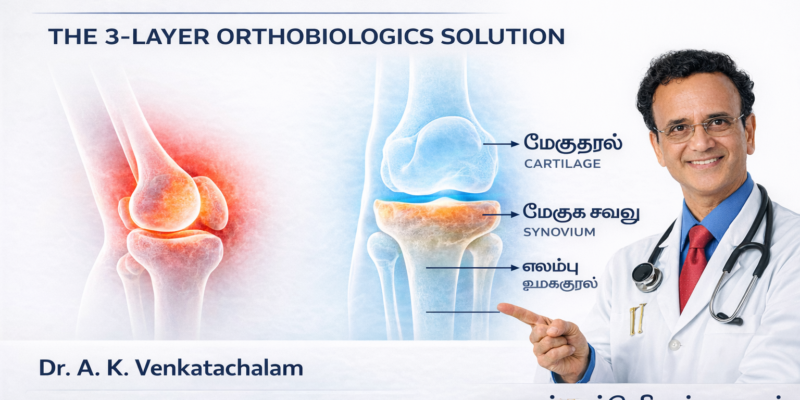
Why Knee Injections Fail — And How the 3-Layer Orthobiologics Approach Solves the Real Problem

By Dr. A. K. Venkatachalam, Madras Joint Rejuvenation Centre

Introduction

Millions of people receive injections for knee pain every year—PRP, hyaluronic acid, steroids, fat injections, “stem cell” treatments, and more.
Yet many patients tell me the same story:

“Doctor, it worked for a while… and then the pain came back.”

Why do knee injections often fail or give only temporary improvement?

The answer lies in a simple but scientifically supported truth:

See the video here

Osteoarthritis is not a single-layer problem.

It is a multi-tissue, multi-layer disease involving:

  1. Synovium – inflammation layer

  2. Cartilage – surface integrity layer

  3. Subchondral bone – load-bearing & pain layer

If you target only one layer, results will be incomplete.
This is where modern Orthobiologics offer a breakthrough—by matching the right biologic to the right biological target.


⭐ The Knee Is a Biological System — Not a Hinge Joint

Recent research describes the knee as a joint organ.
Synovium, cartilage, meniscus, fat pad, and subchondral bone communicate continuously through biochemical, mechanical, and inflammatory pathways.

This means:

  • Inflammation in the synovium affects cartilage.

  • Weakness or edema in the subchondral bone causes nerve sensitization.

  • Cartilage breakdown exposes bone and triggers more inflammation.

So OA progresses not in isolation, but through interconnected tissues.


⭐ Layer 1: The Synovium (Inflammation Layer)

The synovium is the first tissue to flare in many patients.
MRI studies show that synovitis correlates strongly with knee pain—even more than cartilage loss.

Why inject here?

Because reducing synovial inflammation stabilises the joint environment.

Best biologic for this layer:

  • Leukocyte-poor PRP (LP-PRP)
    Evidence shows LP-PRP reduces:

  • IL-1β

  • TNF-α

  • Synovial inflammatory markers

This is why I use LP-PRP as the foundation layer in ageing knees.


⭐ Layer 2: Cartilage (Surface Layer)

Cartilage has poor natural repair capacity due to being:

  • Avascular

  • Aneural

  • Low-cellularity

Orthobiologics do not regrow cartilage in advanced osteoarthritis,
but they can improve:

  • Matrix turnover

  • Chondroprotection

  • Joint lubrication

  • Cellular signalling

Best biologic for this layer:

  • Adipose-derived stromal cells (microfat / SVF-rich preparations)
    These cells support:

  • Matrix maintenance

  • Immunomodulation

  • Cellular cross-talk

In moderate OA, PRP + stromal cells provides stronger and longer symptom relief than PRP alone.


⭐ Layer 3: Subchondral Bone (Load-Bearing Layer)

This is the most overlooked layer, yet it is often the true generator of pain.

Subchondral bone develops:

  • Bone marrow lesions (BMLs)

  • Increased pressure

  • Nerve sensitization

  • Metabolic dysfunction

These changes destabilise the cartilage above.

Why inject here?

Because intraosseous biologics are now shown to:

  • Reduce BMLs

  • Improve load distribution

  • Reduce mechanical pain

  • Support cartilage from below

This is the missing link in many failed treatment attempts.


⭐ Why Injections Fail: The Single-Layer Problem

Most injections target only one of the layers:

Treatment Layer Targeted
Steroid Synovium only
HA Lubrication; minor cartilage effect
PRP Primarily synovium
Fat (microfat) Stromal/cellular layer
BMAC Mixed effects but inconsistent
“Stem cell” injections Usually intra-articular only

If the synovium is treated but the subchondral bone remains diseased, pain returns.
If cartilage signalling improves but synovial inflammation persists, results are weak.

This is why single-injection approaches often fail.


⭐ The 3-Layer Orthobiologic Strategy (My Clinical Framework)

To achieve lasting benefit, each biologic should be chosen according to the target biology:

Layer 1 → Synovium → LP-PRP

Layer 2 → Cartilage → Adipose stromal cells (microfat)

Layer 3 → Subchondral bone → Intraosseous biologics

Not every knee needs all three.
But every knee needs the correct layer addressed.

This biological matching significantly improves outcomes.


⭐ Scientific Support for the 3-Layer Model

Synovium

  • Scanzello CR et al., Bone 2012 — Synovitis drives OA progression

  • Benito et al., Arthritis Rheum 2005 — Pain strongly correlates with synovitis

Cartilage

  • Filardo et al., AJSM 2015 — PRP improves chondrocyte metabolism

  • Buckland J., Nat Rev Rheumatol — MSCs support matrix biology

Subchondral Bone

  • Driban et al., Osteoarthritis Cartilage 2016 — BMLs predict pain & progression

  • Sánchez et al., Int Orthop 2016 — Intraosseous PRP improves BML-related pain


⭐ Who Benefits Most from the 3-Layer Approach?

Ideal candidates include:

  • Men and women 45+

  • With chronic knee pain

  • Early-to-moderate osteoarthritis

  • MRI showing synovitis or BMLs

  • Recurrent pain after PRP or HA

This approach is especially useful in post-menopausal women and metabolically inflamed knees.


⭐ Conclusion

Knee osteoarthritis is not a one-layer problem.
It is a multi-system joint disease, and orthobiologic treatments must be matched to each biological layer:

  • LP-PRP → inflammation

  • Stromal cells → cartilage & signalling

  • Intraosseous biologics → subchondral bone

This 3-Layer model is scientifically grounded, clinically logical, and highly effective for personalized knee preservation.

Q1. Why do knee injections stop working after a few months?

Because they usually treat only one layer (synovium or cartilage) while OA is a multi-layer disease involving synovium, cartilage, and subchondral bone.

Q2. What is the 3-layer orthobiologic approach?

A modern strategy targeting synovial inflammation, cartilage signalling, and subchondral bone remodeling with different biologics.

Q3. Is PRP enough for knee arthritis?

In early OA—sometimes. In moderate OA—usually not. LP-PRP is Layer 1 only.

Q4. Do adipose stromal cells grow new cartilage?

No. They improve signalling, reduce inflammation, and stabilise cartilage microenvironment.

Q5. What is intraosseous PRP?

A targeted biologic injection into the subchondral bone to reduce bone marrow lesions and mechanical pain.

Q6. Who benefits most from this approach?

Patients above 40, especially with synovitis, BMLs, or metabolic knee inflammation.

Metabolic Osteoarthritis

It’s Not Just “Wear and Tear”: The Metabolic Secret Behind Your Chronic Knee Pain

Why standard treatments fail when your own body chemistry is attacking your joints.

For decades, patients have been told a simple story about knee osteoarthritis: it is a disease of “wear and tear.” You used your knees too much, you got older, the cartilage wore down like the tread on a tire, and now it hurts.

But what if I told you that for a significant number of patients, this story is incomplete, or even flat-out wrong?

At the Madras Joint Rejuvenation Centre (MJRC), we see many patients whose pain seems disproportionate to their physical activity or age. They have tried painkillers, braces, and physiotherapy, but the deep, throbbing ache remains.

In my latest video presentation (embedded below), I explain the reason why: we have moved past the simple “wear and tear” model and now understand a distinct, aggressive phenotype known as Metabolic Osteoarthritis.

To understand metabolic arthritis, you must understand inflammation.

Acute inflammation is necessary. If you cut your finger, your body rushes immune cells to the area to fight bacteria and heal the wound. Once healed, the inflammation stops.

In metabolic osteoarthritis, that “stop” signal never comes.

Think of your immune system like a fire alarm in a building. When there is smoke, the alarm blares, and the sprinklers turn on. But imagine if the alarm gets stuck “ON” even after the fire is out. The sprinklers keep running, eventually rotting the wood and destroying the building’s structure.

In your knee, this chronic, low-grade inflammation acts like those sprinklers. It slowly degrades cartilage and irritates the joint lining (synovium), causing persistent pain that mechanical fixes cannot solve.

The Culprit: When Fat Turns Against You

Where does this constant inflammatory signal come from? In metabolic osteoarthritis, it often comes from our own adipose tissue—body fat.

For years, medicine treated fat as inert storage. We now know that adipose tissue acts like an endocrine organ. It is biologically active. Excess fat tissue, particularly visceral fat around the abdomen, secretes powerful inflammatory proteins called adipokines and cytokines.

These chemical messengers travel through your bloodstream and attack vulnerable tissues—including the cartilage in your knees. This is why obesity is linked to arthritis not just because of heavy load-bearing, but because the body is in a state of systemic inflammation.

The “Skinny Fat” Paradox: It’s Not Just About Weight

This is the most crucial point for many patients: You do not have to be morbidly obese to have metabolic arthritis.

In the video, I share a case study of a patient with a normal BMI. Their routine blood tests showed normal fasting glucose. Their doctor told them they were fine.

However, when we ran deeper metabolic panels at MJRC, we found their fasting insulin was dangerously high. Their pancreas was working overtime just to keep blood sugar normal, flooding their body with insulin—a major driver of inflammation.

This patient didn’t need a knee brace; they needed to address their metabolic dysfunction.

Why Conventional Treatments Fail

If the root cause of your knee pain is a metabolic “fire,” you cannot fix it with purely mechanical tools.

  • Painkillers (NSAIDs) temporarily mask the signal but do not stop the fire.

  • Braces provides support but do not stop the chemical attack on the cartilage.

  • Arthroscopic “clean-ups” often fail because the inflammation returns immediately.

The Future: Treating the Chemistry, Not Just the Mechanics

Understanding that osteoarthritis has a metabolic basis is the first step toward real relief. If we can identify that your body is in an inflammatory state, we can change the treatment plan.

At MJRC, we focus on Orthobiologics and protocols like PRASAD™, which are designed not just to patch up the damage, but to alter the biological environment of the joint, helping to turn off that stuck “fire alarm.”

In my next article and video, I will explain exactly how these biologic treatments attempt to influence this inflammatory memory and promote healing.


Are you frustrated with knee pain that won’t resolve? You may be dealing with Metabolic Osteoarthritis. Contact the Madras Joint Rejuvenation Centre today for a comprehensive evaluation that looks beyond just “wear and tear.”

See this link-https://share.google/WgNZniY59jVuxsOu8

Scientific references  for the  video

Scientific References

  • Systemic Inflammation & OA:

    • Berenbaum F. et al. “Osteoarthritis as a systemic disease.” Nature Reviews Rheumatology, 2013.

  • Adipokines & Cartilage:

    • Gualillo O. et al. “Obesity and osteoarthritis: more than just mechanics.” Osteoarthritis and Cartilage, 2007.

    • Conde J. et al. “Adipokines: novel players in rheumatic diseases.” Nature Reviews Rheumatology, 2011.

  • Alarmins & Immune Response:

    • Hotamisligil G.S. “Inflammation and metabolic disorders.” Nature, 2006.

  • Inflammatory Memory:

    • Serhan C.N. “Pro-resolving lipid mediators are leads for resolution physiology.” Nature, 2014.

  • Metabolic Syndrome & Joint Pain:

    • Sellam J., Berenbaum F. “The role of synovitis in pathophysiology and clinical symptoms of osteoarthritis.” Nature Reviews Rheumatology, 2010.

    • Courties A. et al. “Metabolic stress-induced joint inflammation and osteoarthritis.” Nature Reviews Rheumatology, 2015.

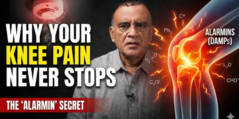
Alarmins, cause of persistent knee pain

Alarmins: The Hidden Reason Your Knee Pain Keeps Flaring Up

Why your knee “screams” even when scans look normal — and how to calm the biology behind it

In This Article, You Will Learn:

🔷 Summary: What You Will Learn

  • What alarmins are
  • Why they trigger sudden knee pain flare-ups
  • How diabetes, insulin resistance, and metabolic stress magnify inflammation
  • Why your MRI can look “normal” while the pain is severe
  • How we treat alarmin-driven knee flares at Madras Joint Rejuvenation Centre (MJRC)


See this video –


🔷 What Are Alarmins?

When your knee joint cells experience stress — from injury, overload, poor metabolism, or inflammation — they release small emergency molecules called alarmins.

Think of alarmins as your joint’s chemical SOS signal.

They alert the immune system:
“Something is wrong — send help immediately!”

Initially, this is protective. But when alarmins stay elevated for too long, the joint becomes stuck in flare-up mode.


🔷 The “Alarmin Cascade”: Why Inflammation Doesn’t Switch Off

The process is simple:

  1. Joint tissues become stressed (overload, metabolic stress, cartilage wear)
  2. Alarmins are released (distress signal)
  3. Immune cells rush in (the joint becomes “hot”)
  4. Inflammation rises suddenly (pain, swelling, stiffness)
  5. Resolution fails (flare lasts longer than expected)

This “failure to switch off” is extremely common in diabetes, obesity, insulin resistance, and metabolic syndrome because their inflammatory baseline is already high.


🔷 Real Case Example

A 62-year-old woman with diabetes came to MJRC with severe knee pain. She had undergone PRS treatment years earlier, and her MRI appeared normal — no meniscal tear, no major cartilage loss.

But her pain suddenly spiked. Her HbA1c was 10.2.

Metabolic imbalance amplified her alarmin response. Once we stabilised her metabolic status and reduced inflammatory load, her pain reduced significantly.

The knee was structurally normal. The biology was not.


🔷 The Metabolic Link: Why Diabetics Have Worse Flares

1. High glucose = more oxidative stress
This irritates cartilage and synovium.

2. Insulin resistance = immune overactivation

Your immune cells become more aggressive.

3. Poor resolution response

Inflammation starts — but doesn’t stop.

This is why many diabetics experience:

  • burning pain

  • night pain

  • sudden swelling

  • pain even at rest

These are chemical flares, not mechanical damage.


🔷 Why Your MRI Can Look “Normal” But You Still Hurt

Most diagnostic scans detect structure, not chemistry.

Alarmins affect:

  • synovial lining

  • cartilage cells

  • immune pathways

But these may not show up on:

  • X-ray

  • MRI

  • ultrasound

So many patients hear:

“Your scan is normal — nothing is wrong.”

Yet their pain continues because the problem is biochemical, not structural.


🔷 How We Diagnose Alarmin-Driven Knee Pain at MJRC

At Madras Joint Rejuvenation Centre, we evaluate both:

  • Mechanical causes (cartilage, meniscus, alignment)

  • Chemical causes (alarmins, metabolism, inflammation)

Our assessment includes:

  • metabolic profile (HbA1c, insulin resistance markers)

  • synovitis evaluation

  • inflammatory triggers

  • mechanical stress evaluation

  • gait and load analysis

This gives a complete picture, not a partial one.


🔷 Treatment: How We Calm Alarmins and Reduce Flares

We follow a stepwise scientific protocol:

1. Reduce metabolic load

  • stabilise blood sugar

  • improve insulin sensitivity

  • lower inflammatory baseline

2. Quiet the alarmin response

  • targeted supplements

  • structured anti-inflammatory nutrition

  • guided low-load exercise

3. Modulate joint biology

Using orthobiologics:

  • PRS (Platelet-Rich Serum)

  • PRASAD protocol

  • BMAC

  • SBE (Subchondral Biologic Enhancement)

These work best when metabolic inflammation is controlled.

4. Restore joint resilience

  • graded strengthening

  • neuromuscular activation

  • metabolic conditioning

This transforms the internal joint environment.


🔷 When Should You Seek Help?

If you experience:

  • knee pain without injury

  • pain at rest

  • sudden flare-ups

  • night pain

  • pain that doesn’t match your MRI findings

  • worsening after sugary meals

  • diabetes + knee pain

…your knee pain may be alarmin-driven, not mechanical.


🔷 Final Takeaway

Your knee isn’t just wearing out — it is signalling.

Alarmins are your joint’s distress signal.
Ignoring them means the cycle continues.
Understanding them gives you control over flare-ups.


🔷 Need a True Biologic Evaluation?

At Madras Joint Rejuvenation Centre, we specialise in evaluating knee pain using:

  • metabolic assessment

  • immune pathway analysis

  • mechanical load evaluation

  • orthobiologic treatment sequencing

If your pain is unpredictable or doesn’t match your scan, you may have alarmin-driven inflammation.

👉 Book a scientific knee evaluation at MJRC.
👉 Let’s calm the biology and restore your joint.

Contact links- E mail- drvenkatjoints@gmail.com

Clinic- https://share.google/if8xFKw1ntV5uby0z

Why Knee Pain Persists: Start Your Recovery by Understanding Inflammation

Chronic knee pain is not just a “wear and tear” problem. It is an active biological process. At the Madras Rejuvenation Centre, our approach begins by helping patients understand why inflammation switches on, why it fails to switch off, and how this disrupts the knee’s natural healing environment.

Your two foundational videos—the Start Here introduction and the Inflammation video—establish the scientific framework for this different way of thinking about knee arthritis.

This article integrates the key concepts from both, giving readers a clear, structured overview of why their knees hurt and how modern biologic medicine now addresses the root mechanisms behind their symptoms.


1. The Real Cause of Knee Arthritis: Failed Inflammatory Resolution

In your Inflammation video, you explain that knee osteoarthritis is not simply a degenerative process. Instead, it represents a state in which inflammation turns on correctly—but fails to resolve.

Inflammation begins for a reason. Even minor stress, micro-injury, or low-grade metabolic imbalance can trigger it. The problem is not the trigger. The problem is the body’s inability to complete the “resolution phase.”

When this resolution fails:

  • Joint lining stays irritated

  • Cartilage remains vulnerable

  • Pain sensors keep firing

  • Synovial fluid becomes less protective

  • Healing switches off

This framework shifts the conversation away from “old age” and toward modifiable biology.


2. Why Inflammation Fails to Switch Off

The Start Here video underscores that modern knee arthritis care must begin with understanding these control mechanisms, not merely suppressing pain.

Here is what we now know:

  • Chronic inflammation is orchestrated, not chaotic.

  • It involves specific molecules, signals, mediators, and checkpoints.

  • When these checkpoints malfunction, the inflammatory programme gets stuck.

Your upcoming sequel videos (Alarmins → Metabolic OA → Biologics → PRASAD) expand on these individual checkpoints.


3. The Two Core Questions Every Patient Must Understand

Your Start Here video frames the patient journey with two essential questions:

Question 1: What keeps turning inflammation on?

This relates to mechanical triggers, metabolic stress, tissue strain, and danger signals inside the joint.

Question 2: Why does inflammation fail to turn off?

This includes disrupted resolution pathways, biochemical imbalances, and molecular “stop signals” that fail.

The Inflammation video gives patients the foundation to explore these deeper mechanisms in a structured, stepwise manner.


4. Why This Matters for Patients

Most patients have only been offered:

  • Painkillers

  • Steroid injections

  • Taping or braces

  • General exercise prescriptions

  • Advice to “lose weight”

But none of this fixes why the joint environment remains inflamed.

Your channel and clinic offer something different: a biologic first approach that restores the natural regulatory system inside the joint—rather than suppressing it artificially.

This empowers patients by:

  • Giving a clear scientific roadmap

  • Explaining the sequence from inflammation → damage → worsening

  • Showing how intervention can be targeted at each checkpoint

Your Start Here video serves as the front door to this roadmap.


5. The MJRC Approach: A Sequential Educational Pathway

Your pillar sequence is designed to build a coherent narrative:

  1. Wear & Tear (myth-busting)

  2. Exercise (benefits and limitations)

  3. Inflammation (foundation)

  4. Start Here (orientation)

  5. Alarmins (danger signals)

  6. Metabolic OA (system-level factors)

  7. Biologic Interventions (restorative tools)

  8. PRASAD (your flagship protocol)

This blog post links your first two steps so patients begin with a correct understanding before accessing deeper content.


6. Watch the Videos for a Clear Starting Point

Start Here Video (Version 1)
Your essential orientation video that outlines the entire philosophy of knee arthritis care at the Madras Rejuvenation Centre.

The Inflammation Video
A foundational explanation of why knee pain persists and what controls the switch of inflammation inside the joint.

These provide the intellectual foundation for patients before entering the more advanced checkpoint videos.


7. What Comes Next

This integrated understanding prepares viewers for your next release:

  • The Alarmins video: the molecules that keep inflammation switched on

  • Followed by Metabolic OA, Biologics, and PRASAD treatment

Any patient entering your ecosystem with this two-video primer will now understand:

  • Why knee arthritis is a biological disorder

  • Why it progresses

  • Why conventional treatments often fail

  • Why your system focuses on resolution instead of suppression

The Scientific Evidence Behind Inflammation-First Knee Arthritis Care

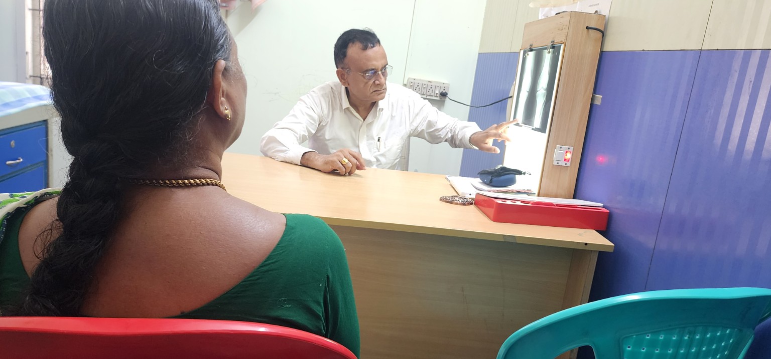
Dr. Venkatachalam discussing radiographic findings during a clinical consultation
Imaging is interpreted alongside the clinical assessment. The photograph illustrates the consultation process, rather than a treatment result.
  1. Scientific Basis of Inflammation-First Knee Arthritis Care

This page summarises the scientific evidence supporting an inflammation-first approach to knee osteoarthritis, as presented in the Inflammation Pillar Video. Each section corresponds directly to a key statement from the script, supported by peer-reviewed research.

1. Knee Arthritis Is Not Just Wear and Tear

Scientific Proof

Osteoarthritis is now recognised as a whole-joint inflammatory disease involving the synovium, cartilage, subchondral bone, ligaments, and infrapatellar fat pad.

Key References

  • Robinson WH et al., Nature Reviews Rheumatology – Osteoarthritis as an inflammatory disease
  • Berenbaum F, Osteoarthritis and Cartilage – Low-grade chronic inflammation in OA progression

Consensus

Mechanical damage alone does not explain pain severity or disease progression.


2. Inflammation Inside the Joint Drives Pain, Stiffness, and Progression

Scientific Proof

  • Synovitis strongly correlates with pain severity
  • Associated with effusion and stiffness
  • Predicts faster cartilage loss

Key References

  • Hill CL et al., Annals of the Rheumatic Diseases
  • Felson DT et al., Arthritis & Rheumatology

Consensus

Pain tracks inflammatory activity, not X-ray grade.


3. Two Patients With the Same X-Ray Can Have Very Different Pain

Scientific Proof

Radiographic severity correlates poorly with clinical symptoms.

Key References

  • Bedson J, Croft PR, Rheumatology
  • Hannan MT et al., Arthritis & Rheumatism

Consensus

Imaging underestimates biological disease activity.


4. Exercise Improves Support, Not Inflammation

Scientific Proof

  • Improves muscle strength and neuromuscular control
  • Does not suppress synovial cytokines when inflammation is active

Key References

  • Henriksen M et al., Osteoarthritis and Cartilage
  • Baker KR et al., Arthritis Care & Research

Consensus

Exercise is necessary but biologically insufficient when inflammation is uncontrolled.


5. Why Patients Plateau Despite Good Physiotherapy

Scientific Proof

  • Active synovitis predicts poor rehabilitation response
  • Associated with pain flares after loading
  • Reduces tolerance to strengthening

Key References

  • Scanzello CR et al., Clinical Orthopaedics and Related Research
  • Schaible HG, Nature Reviews Rheumatology

Consensus

Plateaus are biological, not motivational failures.


6. Painkillers Suppress Symptoms, Not Disease Biology

Scientific Proof

  • NSAIDs reduce pain temporarily
  • Do not halt cartilage degeneration
  • Do not modify disease progression

Key References

  • Zhang W et al., OARSI Guidelines
  • Hochberg MC et al., Arthritis Care & Research

Consensus

Symptom relief does not equal disease control.


7. Sequence Matters: Calm Inflammation Before Strengthening

Scientific Proof

  • Reducing inflammatory load improves pain thresholds
  • Improves exercise tolerance
  • Enhances functional outcomes

Key References

  • Atukorala I et al., Arthritis Research & Therapy
  • Bennell KL et al., British Journal of Sports Medicine

Consensus

Biological readiness determines rehabilitation success.


8. Joint Preservation Focuses on Biology, Not Just Mechanics

Scientific Proof

Modern OA management emphasises early biological modulation, load management, and individualised treatment sequencing.

Key References

  • Loeser RF et al., Osteoarthritis and Cartilage
  • Hunter DJ, The Lancet

Consensus

Joint preservation is proactive, not passive.


9. Inflammation Is Treatable

Scientific Proof

  • Inflammation in OA is measurable
  • Inflammation is modifiable
  • Inflammation is clinically meaningful

Key References

  • Mathiessen A, Conaghan PG, Arthritis Research & Therapy
  • Scanzello CR, Goldring SR, Arthritis & Rheumatology

Consensus

Osteoarthritis inflammation is manageable, not inevitable.

See the video here -https://youtu.be/IJFPdKUwpc0?si=5lN4781-zEmo3Xk0

FAQ’s

Q: Is knee osteoarthritis only caused by wear and tear?

A: No. Current research shows osteoarthritis is a whole-joint inflammatory disease. Mechanical wear alone does not explain pain severity or progression.

Q: Why does knee pain not match X-ray findings?

A: Pain correlates more strongly with synovial inflammation than with radiographic cartilage loss. X-rays underestimate biological disease activity.

Q: Does exercise reduce inflammation in knee arthritis?

A: Exercise improves strength and support but does not reliably suppress active synovial inflammation when it is present.

Q: Why do some patients plateau despite physiotherapy?

A: Active inflammation sensitises pain pathways and limits tolerance to loading, leading to biological—not motivational—plateaus.

Q: Are painkillers disease-modifying in knee arthritis?

A: No. NSAIDs reduce symptoms temporarily but do not alter the underlying disease process or progression.

PRASAD Treatment for Knee Osteoarthritis: What It Is, Who It Helps, and How It Works

  1. PRASAD Treatment for Knee Osteoarthritis: What It Is, Who It Helps, and How It Works

Most people believe knee osteoarthritis is simply a problem of worn-out cartilage. Modern research, however, shows that osteoarthritis is driven by chronic inflammation, metabolic imbalance, and altered cartilage cell behaviour.

The PRASAD Treatment was developed to address these deeper mechanisms. Rather than offering temporary pain relief, it aims to reset the joint’s biological environment so that function improves and disease progression slows.

Why Osteoarthritis Needs More Than Symptom Control

Osteoarthritis is now understood as a whole-joint disease involving cartilage, synovium, bone, muscles, and inflammatory signalling molecules. Persistent inflammation keeps the joint in a breakdown-dominant state, even when X-rays appear only mildly abnormal.

This explains why pain severity often does not correlate with imaging findings—and why isolated injections or painkillers rarely provide durable improvement.

What Is the PRASAD Treatment?

PRASAD is a structured regenerative protocol designed to:

  • Reset chronic joint inflammation
  • Improve cartilage and synovial metabolism
  • Enhance the joint’s internal repair environment
  • Restore strength-based load tolerance
  • Delay or avoid knee replacement in selected patients

Unlike single-shot therapies, PRASAD is a phased protocol tailored to the patient’s inflammatory status, metabolic profile, and stage of osteoarthritis.

The Biological Rationale Behind PRASAD

Inflammation Reset

Stressed cartilage cells release danger signals known as alarmins. These molecules perpetuate inflammation and accelerate tissue breakdown. PRASAD targets this inflammatory loop to calm the joint before regenerative stimulation is applied.

Metabolic Rebalancing

In osteoarthritis, cartilage cells shift toward a catabolic (breakdown-driven) state. PRASAD incorporates metabolic correction and biologic modulation to push the joint environment back toward repair and stability.

Targeted Regenerative Support

Depending on patient selection, PRASAD may integrate platelet-based or cell-supported biologics, always within a controlled protocol rather than as stand-alone injections.

How PRASAD Differs From Standard Knee Injections

Common Treatment Primary Action PRASAD Difference
Steroid injections Short-term inflammation suppression PRASAD avoids cartilage-weakening effects of repeated steroids
PRP Growth factor delivery Used only after inflammation is biologically controlled
Hyaluronic acid Lubrication PRASAD focuses on biological reset, not temporary viscosity

Who Is an Ideal Candidate?

PRASAD is most effective for patients with:

  • Early to moderate knee osteoarthritis
  • Inflammatory flares with activity-related pain
  • Stiffness that improves with movement
  • Metabolic risk factors such as weight gain or insulin resistance
  • A desire to delay or avoid knee replacement

Expected Outcomes

Patients commonly experience improvements in pain, swelling frequency, walking endurance, stair climbing, and confidence in knee function. Results are gradual but more durable because the underlying joint environment is altered.

Safety Considerations

PRASAD primarily uses autologous biologics combined with structured rehabilitation and metabolic correction. This results in a favourable safety profile when proper screening is followed.

Why PRASAD Represents the Future of Osteoarthritis Care

Modern osteoarthritis management is moving toward early biologic intervention, inflammation control, metabolic optimisation, and strength-based joint loading. PRASAD aligns with this evidence-driven direction.

For a simpler, patient-focused explanation of the PRASAD approach, read the detailed overview on drakvenkat.com.

Long-Term Follow-Up Update (2026)

We recently received a follow-up review from a patient who underwent our stromal-based biologic treatment 6 years ago.

They continue to report:

Sustained pain relief

Stable knee function

See the attached screen shots

Active daily lifestyle6 year follow up of PRS treatment

No need for knee replacement

  1. This long-term outcome reinforces the durability of biologic knee-preservation strategies when applied to appropriately selected patients.

Continue reading “PRASAD Treatment for Knee Osteoarthritis: What It Is, Who It Helps, and How It Works” →