
For about five years, a patient from Canada lived with knee osteoarthritis that made walking longer distances and negotiating stairs increasingly difficult. Painkillers, ointments, steroid injections and hyaluronic acid had provided relief for only a few months at a time. He was looking for options that might help him defer knee replacement.
After finding the clinic’s website, he contacted Dr. Venkatachalam by email. The correspondence and educational material helped him understand the proposed joint-preserving approach. He travelled to Chennai for assessment and treatment.
Assessment and treatment
His assessment showed medial-compartment osteoarthritis, visible bowing, difficulty fully straightening the knee and patellofemoral symptoms. He underwent PRASAD-based orthobiologic treatment, followed by rehabilitation and the advised use of patellar and unloading support.
Follow-up
Recovery took time. At around three months, he reported that pain had settled, although going down stairs still caused some discomfort. Over the following months, his walking and stair use continued to improve. At around 10–11 months, he reported walking more comfortably and being able to squat fully for about two months and sit cross-legged. He was using his brace less often indoors and continued strengthening and balance exercises. At follow-up, the standing appearance of the knees seemed less bowed than in earlier photographs. This is a visual observation only; no standardized measurement was provided, so it does not establish a change in underlying bone alignment.
At follow-up
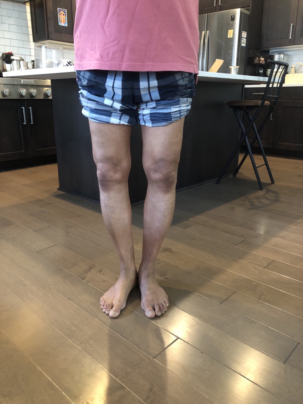
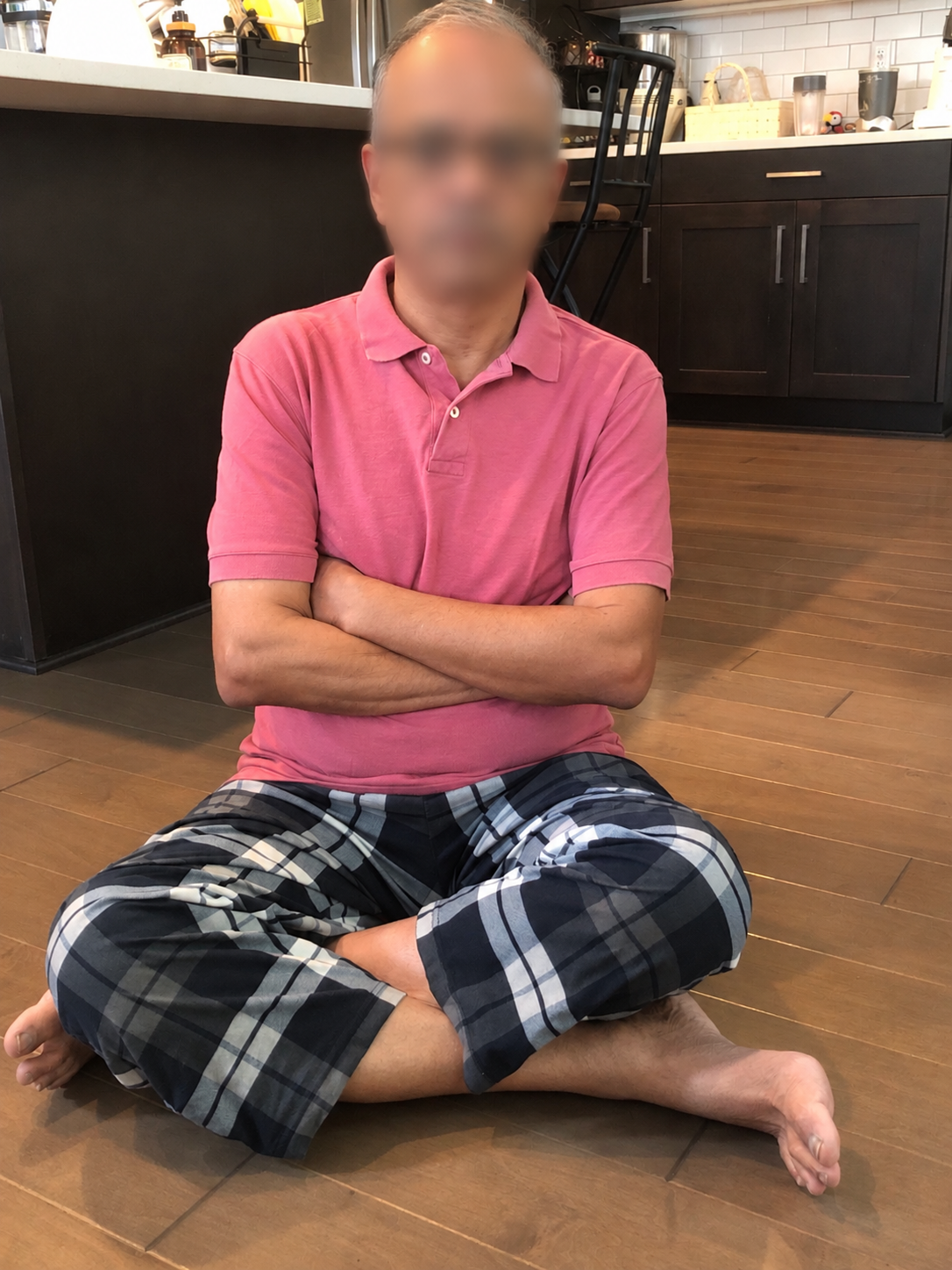
This account combines the patient’s own experience with follow-up observations. It describes one person’s course after a combined treatment and rehabilitation programme; it cannot establish which component produced the change or predict another person’s outcome. The photographs show function and appearance, not cartilage regeneration.
Related reading: Bone marrow lesions
For general background on bone marrow lesions (BMLs) in knee osteoarthritis and research on the bone beneath cartilage, read The Bone Beneath the Cartilage: What the Research Shows. This is general evidence context; it does not indicate that this patient had a BML.